Ask For A Quick Quote
We will contact you within 1 working day, please pay attention to the email with the suffix “@fsdym.com”.
We will contact you within 1 working day, please pay attention to the email with the suffix “@fsdym.com”.
Working Principle of the Oral Endoscope
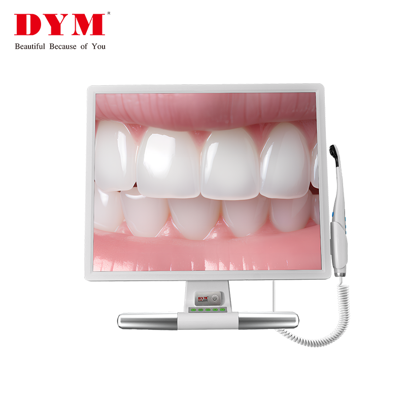
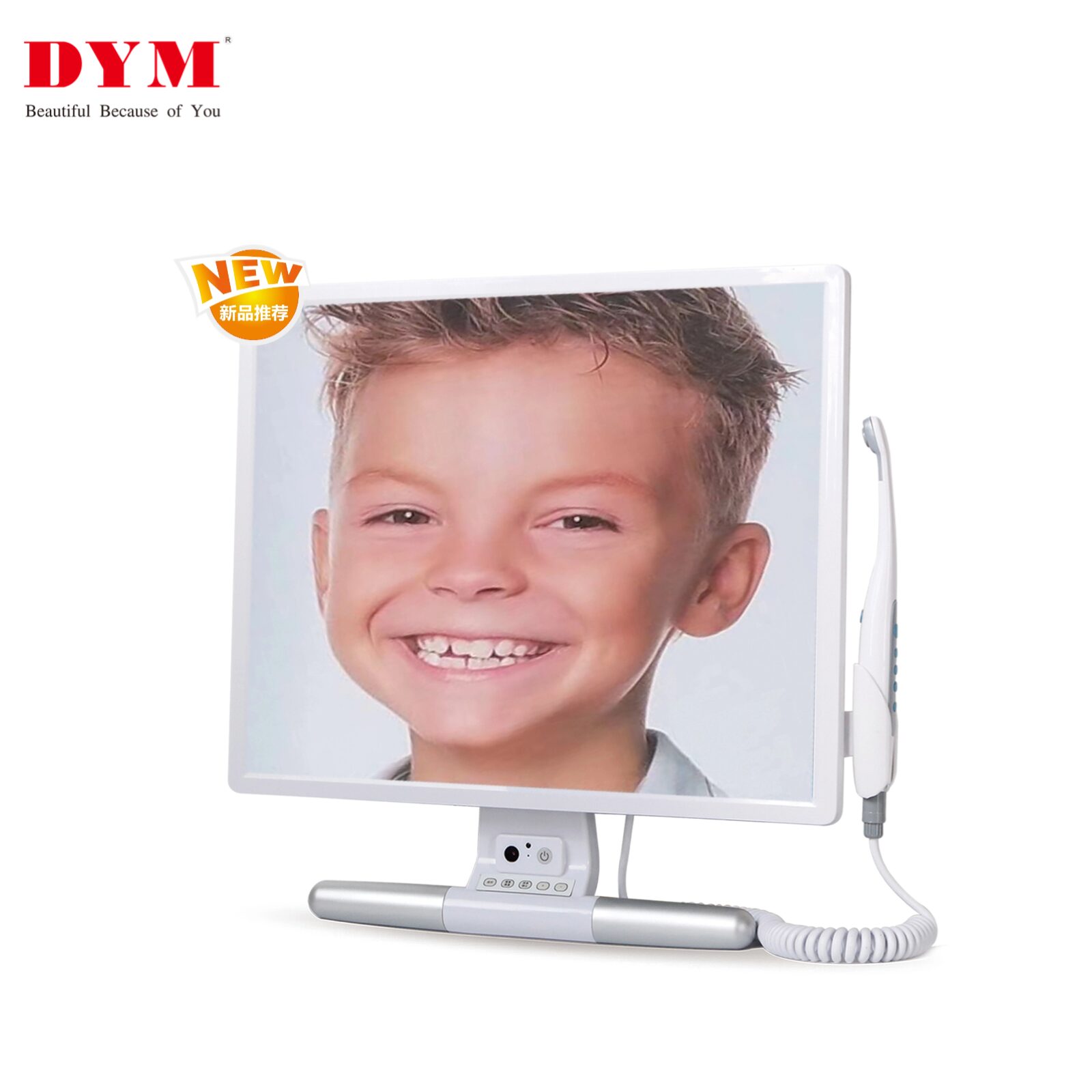
The oral camera typically consists of a display monitor, a handheld device, and a support bracket.
Optical Principle of the Handheld Unit:
The front end of the oral camera is equipped with an optical lens and a light source. It utilizes the principle of total internal reflection to transmit images from inside the oral cavity as digital signals to the monitor. This allows the dentist to clearly observe the condition inside the mouth.
Imaging Principle:
A miniature camera captures images within the mouth and converts them into electrical or digital signals, which are then transmitted to a connected high-definition screen. This real-time display clearly shows the teeth, gums, and mucosal surfaces inside the oral cavity.

Components of the Oral Endoscope
Camera Probe:
The front part of the oral camera is typically slim and elongated, allowing it to reach all corners of the oral cavity. The probe tip contains a miniature camera and light source—the camera captures images, while the light provides sufficient illumination for clear visuals.
Handheld Connector:
This part connects the probe to the display and is designed for easy handling by the dentist. It usually features control buttons for adjusting the camera’s focus, direction, brightness, and may also include functions such as image capture, video recording, and storage.
Display Monitor:
Used to display real-time images captured by the probe. Typically a high-definition screen, it presents clear and accurate details of the oral cavity, allowing both doctors and patients to see the internal condition visually.

Advantages of DYM Oral Camera
High-Definition Imaging:
Provides real-time, high-resolution images that clearly show the fine structures and lesions in the teeth, gums, and oral mucosa, aiding accurate diagnosis.
Non-Invasive Examination:
Compared with traditional oral examination methods, the oral Camera offers a non-invasive approach that causes no damage to oral tissues, with minimal discomfort—making it easier for patients to accept.
User-Friendly Operation:
Human-centered design allows for easy operation. The dentist can flexibly adjust the probe’s position and angle to observe different parts of the mouth comfortably, while patients can maintain a relaxed posture during the exam.
Diagnostic and Treatment Aid:
Helps dentists detect potential oral diseases and provides real-time visual guidance during treatments such as dental implants, root canal therapy, and periodontal surgery—improving precision and success rates.
Patient Education:
Patients can see the inside of their mouth in real time via the monitor, giving them a better understanding of their oral health and existing issues. This enhances their trust in the doctor’s diagnosis and treatment plan and improves treatment outcomes.
DYM Oral Camera Models:
Currently available in four models: SKI-102, SKI-103, SKI-105, and SKI-106.
SKI-103 features:
19-inch display screen
1380-pixel resolution
Original BOE high-definition screen
Built-in camera for recording doctor-patient interactions with playback function
Rewatchable video footage
Disinfectable aluminum alloy handle for better hygiene
Aviation-grade flexible and durable cable
16GB original USB flash drive capable of storing approx. 2000 images
Warranty & Lifespan:
1-year warranty and an extended 5-year effective usage lifespan for your peace of mind.